Surgeons are trained to utilize various surgical procedures in order to physically reach inside of an individual’s body in order to diagnose or treat medical conditions. Surgery procedures may be lifesaving, corrective or cosmetic in nature.
Reconstructive surgery on specific parts of the body usually begins with a prefix to specify where exactly it will take place, for instance, “rhinoplasty” (nose reconstruction). Lister’s pioneering efforts in developing sterile surgery practices with steam sterilizers, antiseptics and rigorous hand washing served as the foundation of modern invasive techniques.
Removing Cancer
Surgery to remove cancerous cells from a specific part of your body is the standard treatment for many forms of cancer, either alone or combined with radiation therapy or chemotherapy. Surgery may even provide a permanent cure if your cancer is small and limited to its original site of origin (primary), as well as lower the chances that it will return (recur).
Docs often remove some healthy tissue around tumors to help prevent cancer from coming back and reduce side effects, like pain. Staging procedures involve extracting lymph nodes near cancerous tumors for analysis; this helps doctors assess how far spread it has become. Based on results of surgery and tests, they will then recommend the best course of treatment.
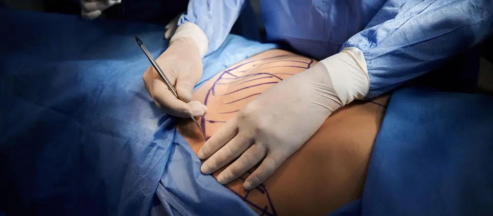
Your surgeon will make an incision, or cut, where the cancerous tissue resides to remove it. They may use special tools or devices like lasers to cut away cancer and seal blood vessels near it so as to minimize bleeding during and after the operation.
A surgical margin refers to the healthy tissue surrounding a tumor. Under a microscope, pathologists examine this tissue for any cancer cells present; if none exist then this area of healthy tissue is known as clear, negative, or clean margin. If however, tumor cells have spread past this area then further treatment may be required.
Preventive surgery can help lower your risks of cancer. This might involve having polyps removed before they develop into cancerous polyps. Or it might involve having breast or ovarian removal if there is an increased family history or mutations to either BRCA1 or BRCA2 genes, making you more prone to getting them.
Lung Cancer Surgery
Your doctor will recommend lung cancer surgery depending on its size, location and stage as well as how well your lungs function. In some instances, when detected early it may be possible to only remove affected portions of lung – this allows for normal breathing while remaining cancer-free. Your surgeon will use different techniques suited to you situation or cancer type to accomplish this feat.
If the cancerous tumor lies near where airways enter the lungs, your doctor may perform a lobectomy procedure to remove it. Lungs consist of parts called lobes; three on the right lung and two on the left; your surgeon will surgically extract both the affected lobe and any surrounding lymph nodes. If, however, your cancer has spread beyond one lobe, your physician might perform a pneumonectomy instead – this means extracting all or part of one lung when other methods have failed in getting rid of your tumor(s).
Your surgeon may use keyhole surgery (also known as video-assisted thoracoscopic surgery (VATS) to perform lung cancer operations without making large cuts in your chest. In this process, doctors make small incisions on either side of your chest and pass a tube containing tiny cameras and operating instruments through them to monitor what they’re doing – leading to reduced hospital stays and faster recoveries.
Some doctors also employ VATS surgery for more complex operations. This technique, similar to non-small cell cancer operations, is usually utilized when hospital stays last 3-7 days or more.
Surgeons will perform what is known as a sleeve resection if lung cancer has spread into one of its airway tubes (bronchi). Part of the bronchus and surrounding lung lobe will be removed before rejoining healthy ends together again afterwards. They might also take measures such as checking nearby lymph nodes for any sign of further spread of tumour cells.
Thoracoscopy
Thoracoscopy is a minimally invasive medical procedure that enables doctors to directly examine the inside of the chest cavity and lungs. Additionally, this minimally invasive process is often used to drain pleural fluid or perform biopsy and wedge resection procedures for various conditions. Pulmonologists typically perform medical thoracoscopy under general anaesthesia or with local anaesthesia for additional comfort.
Before having thoracoscopy, imaging studies such as CT scan, chest X-ray or electrocardiogram will likely be necessary depending on your type of procedure. Your healthcare team will explain these tests to you in advance and tell you what to expect during them; in some instances you may even need to stop taking certain medications or supplements prior to receiving this procedure.
Doctors make small cuts (incisions) in your ribcage to insert a thin, lighted tube with a camera called a “thoracoscope (pronounced “thuh-RAY-kuh-skohp”) into your chest cavity and lungs for examination. They use this instrument to view both spaces simultaneously; should any additional instruments need be introduced through other incisions within that same incision, the doctor can insert other tools to perform surgical procedures like taking biopsies or draining fluid from chest cavity cavities or draining chest cavities.
Though thoracoscopy carries some risks, in most cases there are no complications. Some individuals may feel some numbness in the area where a cut was made but this should subside with time. You may need a tube in your chest for two days afterward to drain fluid out.
Some patients undergoing thoracoscopy may encounter the complication known as tumor seeding, in which cancerous cells spread to areas beyond those targeted by surgery. Radiation therapy can help avoid this complication.
After your thoracoscopy procedure is over, your physician will go over its results and discuss any necessary treatments or follow-up care with you. In most cases, you should be able to go home on the same day or within days depending on what was discovered; if a biopsy was taken then this may take some additional time; alternatively a lab may need time to send its report with results back.
RATS
Rats may have an unfavorable reputation as vermin, but they make rewarding pets. Rats form strong bonds with their caretakers, are highly intelligent creatures with complex needs that may prove challenging to meet; nocturnal in activity at dawn and dusk with excellent hearing capabilities as well as superior senses for scent detection.
These creatures typically feature long bodies with narrow, pointed heads. Large eyes and prominent ears often accompany this trait, along with short tails relative to body length that may appear lighter in color than their coat. Their very powerful sense of smell allows for them to have long chisel-shaped incisors; their paws are narrow and usually bald; some species feature long guard hairs which give tufted features around ears and tail.
Rats are polygynandrous animals, meaning both females and males in a group have multiple partners. Rats reach sexual maturity around four months old and typically produce seven litters annually – typically breeding between spring and autumn but some tropical species breed all year-long – the gestation period being 21-26 days.
House rats tend to be omnivorous eaters that consume almost anything digestible – particularly stored grains – while brown rats from forests and woodland areas can be more carnivorous, hunting small vertebrates such as shrimps, snails, mussels, insects, amphibians, bird eggs/young and poultry as prey items.
Some rat species possess very short, dense coats with soft fur while others have longer, coarser ones. Himalayan field rats (Rattus norvegicus) feature bicolored tails whereby their upper surface is brown or dark gray while their undersurface has paler tones; Hoogerwerf’s Rat of Sulawesi and Sikkim White-tailed Rat have brown all over except for basal third to half of tail, which remains pure white.
The Smithsonian’s National Zoo feeds its rat population primarily with rodent block diet, supplemented with fresh vegetables and fruits for enrichment. Our enclosures are clean, well-lit, temperature-controlled environments. Furthermore, trained staff handle them on an ongoing basis.
Disclaimer: The content on this blog is intended for general informational purposes only. It is not a substitute for professional medical advice, diagnosis, or treatment. Always consult qualified healthcare providers for personalized advice. Information regarding plastic surgery, dental treatment, hair transplant, and other medical procedures is educational and not a guarantee of results. We do not assume liability for actions taken based on blog content. Medical knowledge evolves; verify information and consult professionals. External links do not imply endorsement. By using this blog, you agree to these terms.


