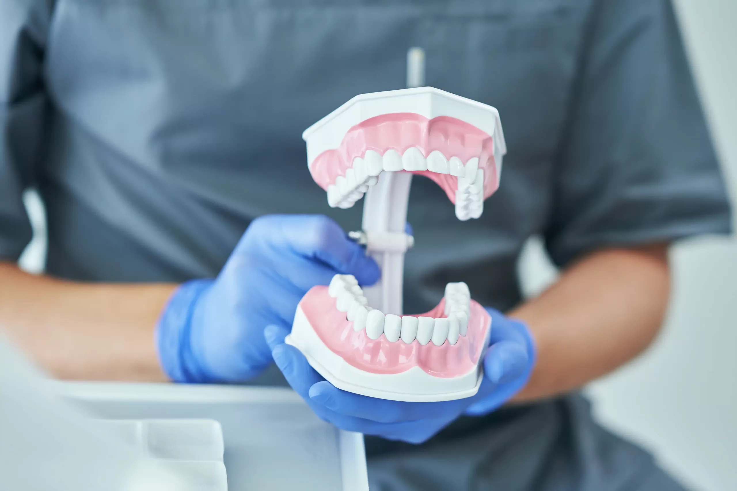
The jaw is composed of bones and muscles that work together to enable chewing, speaking, and breathing. It includes both an upper fixed part (maxilla) and a moveable lower part (mandible).
Habits that stress out jaw muscles and joints include clenching or grinding teeth, biting nails or pencils, touching or resting your cheek or lip frequently and opening wide for extended periods such as when yawning.
Bones
The jaw is composed of two bones: the maxilla (upper jaw) and mandible (lower jaw). Both of these structures contain numerous muscles, nerves and blood vessels that help hold teeth in place as well as support soft tissue structures like tongue and lips. Together these bones work to open and close your mouth – with two TMJs connected at either side allowing it to move up and down, side to side and forward and backward in relation to skull.
The mandible is the larger of two bones, supporting the lower part of the face. Its wings, or ramus, extend out from its body of jaw and meet at two ridges: coronoid process in front, condylaris process at back; these both bind the temporomandibular joint. In addition, its ramus has several openings (fossae) which allow essential nerves and arteries to access mouth region.
These include the mental foramen, which allows the mental nerve to travel from the chin to the lower part of the jaw; an incisive fossa for receiving the incisor and canine teeth; and finally the palatine foramen which is located in the upper portion of the palate and provides passage for the palatine molars – as well as providing passageway for inferior salivary nerve from ramus to body of jaw via an oblique line.
Both upper and lower jaws contain many other crucial openings. They hold salivary glands, which produce fluid to moisten food and drink, food entering and exiting through openings in the lower jaw, the lingual nerve (only one felt in tongue), which provides sensation to lower part of tongue as well as small part of cheek, as well as the largest and strongest teeth known as molars – designed specifically to grind food for swallowing or chewing purposes – found there as well.
Muscles
Muscles in the jaw are responsible for opening and closing it by manipulating motion of the lower jaw bone (mandible). These muscles can generate up to 200 pounds of pressure during chewing and speaking – known as the masticatory or chewing muscles. Four muscles play a part in this mastication: masseter, temporalis, medial pterygoid, and lateral pterygoid muscles are involved; these are further divided into superficial groups consisting of masseter and temporalis muscles while deep groups that include medial pterygoid muscles (masseter and temporalis), medial Pterygoid muscles).
The masseter muscle is one of the strongest of masticatory muscles, consisting of four quadrangular layers separated into deep and superficial sections by horizontal fibres that connect anterior fibres vertically while middle sections have an oblique orientation, and posterior ones horizontal. It serves to protract and elevate jaw closure during chewing processes.
Masseter muscles are one of four involved in mastication. This powerful muscle in your body contracts when contracted to cause powerful elevation of the mandible and shutting of your mouth. Due to its insertion along angle and lateral surface of ramus it also plays a vital role during protrusion of jaw during opening of mouth.
The temporalis muscle is a fan-shaped muscle located in the lateral temporal fossa of the skull that comprises anterior, middle, and posterior fibres that lie in an oblique and horizontal orientation. It serves to raise and lower the mandible during mastication as well as contribute to side-to-side grinding movement of teeth. Its supply comes from deep temporal nerve and medial branch of maxillary artery.
The lateral pterygoid muscle is a triangular structure composed of two heads: superior and inferior. The superior head contains vertically aligned muscle fibers to retract the lower jaw while its inferior head features horizontally-oriented fibers which protrude it, functioning to protrude it as it lies below medial pterygoid muscle.
Joints
Joints connect two bones and allow movement in your skeleton. There are numerous different kinds of joints; some may allow back and sideways movement while others can rotate freely; the human skeleton contains over 260 joints, some being rigid enough that only slight angles of movement are possible.
Joints are composed of the intersection between two bones joined together with cartilage or ligaments, held together by their ends. Some joints may even be lined with synovial fluid and known as synovial or gliding joints. Bones can move through these joints in different ways called articulations which results in movements such as flexion, extension, protraction retraction elevation depression etc.
Some joints do not move at all and are known as immovable or fibrous joints; such as sutures in the skull and the articulations between teeth and mandible or maxilla. Others are only capable of minor movement and are known as movable diarthroses joints.
Joints are classified by both their structure and function. Structural classification identifies bony, fibrous, cartilaginous joints; while functional classification classifies them by specific movements such as angular rotational or special movements.
Vertebrate animals’ movable jaws consist of two vertical sections, including a lower jaw (mandible) that moves and an upper jaw that remains fixed (maxilla). Both parts of the jaws are connected by Meckel’s cartilage which forms a fibrocartilage joint between it and bones in their neck and face regions, providing stability during biting and chewing functions. These parts work in concert to perform these essential actions of chewing.
Some movable jaws are less flexible than they seem and are considered rigid or immovable. Some are composed of bony plates which fusion after birth or shortly thereafter and form gomphoses at their intersections with roots of teeth and jaw bones, or fibrous connections between these roots and bones; other jaws may be created from cranial plates as well as from their connections with skull ribs, pelvic girdle, or pelvic floor muscles.
Function
The jaws are one of the most complex joints in the human body. Their complex joint architecture makes them essential in chewing, swallowing, speaking and even yawning – and when working optimally they allow these actions to occur smoothly and coordinated – otherwise known as Temporomandibular Disorders or TMD. When not functioning optimally this leads to TMD as it connects the lower jaw bone (mandible) to temporal bones on both sides of the head through two joints called TMJs in front of each ear which connect lower jaw bone (mandible) directly. Muscle control over these joints allows movement up or down, side to side as well as forward/back.
The jaw’s articulation plays an essential role in food movement and digestion, forming the roof of the mouth as well as fulfilling other vital roles. Furthermore, its movement impacts facial expressions dramatically while contributing to respiratory, digestive and endocrine health.
Temporomandibular joints play an integral role in both behavioral and emotional responses. They serve as the site for powerful masseter muscle, responsible for chewing activities. Furthermore, this joint serves as the focal point for cranial nerve which provides motor function and sensory input to jaw and tooth muscles through trigeminal nerve bundles that pass through it.
Due to this intimate connection between injuries to jaw bones or temporomandibular joints and ears, injuries sustained by either can affect them directly. When people report experiencing earache or jaw pain they often attribute it to dental problems when in reality it could be the TMJ at work.
Function of the jaws depends upon a number of factors, most importantly those related to TMJ (Temporomandibular Joint), muscles, ligaments, disk and teeth. Therefore, having an ideal bite is not only key for optimal functionality but also essential in maintaining both health and appearance of teeth and gums.
Disclaimer: The content on this blog is intended for general informational purposes only. It is not a substitute for professional medical advice, diagnosis, or treatment. Always consult qualified healthcare providers for personalized advice. Information regarding plastic surgery, dental treatment, hair transplant, and other medical procedures is educational and not a guarantee of results. We do not assume liability for actions taken based on blog content. Medical knowledge evolves; verify information and consult professionals. External links do not imply endorsement. By using this blog, you agree to these terms.


