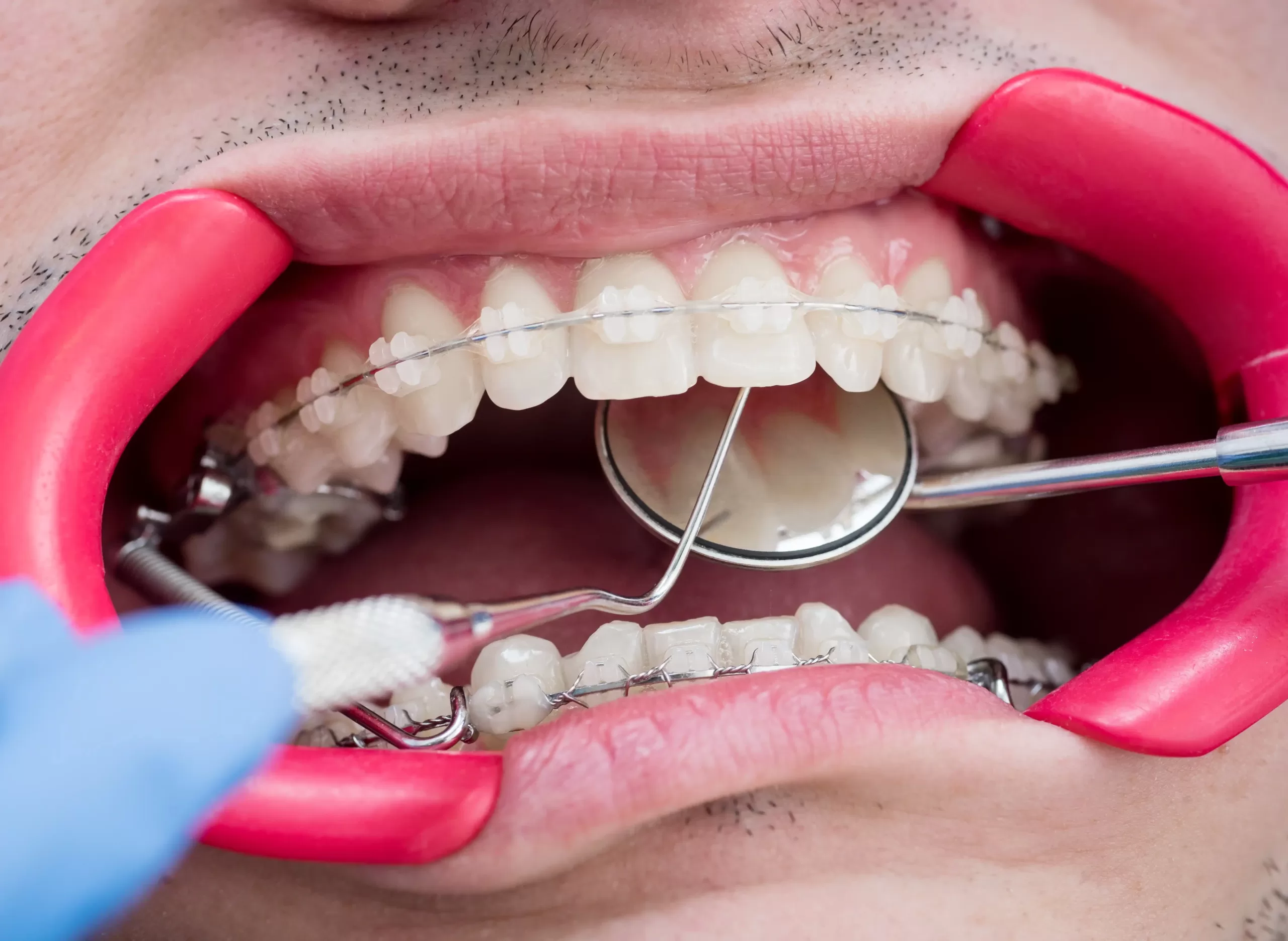
Enamel is the hardened outer surface of your teeth and provides protection from bacteria as well as aiding them to withstand pressure during chewing.
An enamel hole, commonly referred to as a cavity, allows acid and bacteria to seep into the soft dentin beneath. Over time this may damage your tooth irreparably.
Enamel
Tooth enamel is a semitranslucent covering on the crown (the part visible outside your mouth) of your teeth that provides protection for dentin and cementum beneath. It serves as the first line of defense against decay; any wear to this protective shell due to genetics, poor brushing habits, acidic foods/drinks/GERD or chronic sinus issues could result in cavities and severe tooth pain; once lost it can’t grow back. Therefore it is crucial that good oral care and healthy eating habits be put in place in order to preserve this protective shell against potential risks!
Tooth enamel consists of a dense concentration of minerals that make up 95% of its composition, making it stronger than bone yet thinner than most vertebrate bodies. Due to a lack of nerves or blood vessels in enamel’s composition, its strength lies within itself – as its hardness rivals bone.
Researchers are continuing their exploration of this complex material. Stefan Habelitz of the UCSF School of Dentistry has been using microscopy techniques to observe how human enamel develops over time, using microscopic images. He discovered that proteins present in enamel align in columns which bend on a molecular level in order to absorb vibrations – helping protect other parts of your teeth from sudden shocks like eating hard candy or experiencing sudden temperature change from eating ice cream.
At every meal and beverage you consume, bacterial acids that process sugars in your mouth produce acidic substances that attack tooth enamel if exposed to acid-rich foods and beverages, leading to tooth decay characterized by sensitivity, discoloration, cracks, or twinges of pain when you eat hot or cold items as well as cupping that exposes dentin. This process is known as tooth decay; signs that can help detect it include sensitivity, discoloration, cracks or cupping that expose dentin.
Dentin
Dentin is a yellow-hued, porous and mineralized tissue composed of cells called odontoblasts that lie beneath enamel in both crown and root regions, where it’s protected by cementum. If an enamel wears down it exposes dentin which then becomes vulnerable to bacteria and decay – hence why practicing good oral hygiene through brushing, flossing and regular dental visits are so vitally important.
Dentin, like enamel, is a tough material. However, unlike bone or cementum, dentin has less hardness and density compared to these materials and also contains microtubules which run to its pulp chamber containing blood and nerve tissues; these tubules help a tooth sense hot or cold temperatures as well as acidic or sticky foods.
Dentin is less mineralized than enamel (96% in an ideal tooth), yet more mineralized than cementum (65-7%). Dentin comprises 70-72% inorganic materials such as crystalline hydroxylapatite and noncrystalline amorphous calcium phosphate crystals; 20% organic material such as collagen type 1, which makes up roughly half the protein content; and 8-10% water.
At present, it appears that different parts of dentin possess unique chemical composition, although its cause remains elusive. Intertubular dentin is filled with protein while peritubular dentin contains none; some experts also speculate that its composition differs depending on whether or not odontoblasts were present during formation.
Dens in Dente (also called dens in dente or “tooth within a tooth”) occurs when enamel folds over into dentin during tooth development, creating an “extra tooth.” If left untreated, this condition leaves teeth vulnerable to infection and decay – therefore seeking professional diagnosis and treatment, including root canal therapy is recommended.
Cementum
Cementum is a hard connective tissue that covers the root surfaces of teeth and securely joins them to their dentin. Light yellow in color, this tissue covers each root surface of each tooth while joining them together firmly with dentin. Thinner near the neck and thicker at root apex, cementum contains mineralized (mainly apatite) and organic materials such as glycoproteins and collagen, in addition to water; its cells responsible are called cementoblasts.
As with enamel, cementum is extremely resilient. Chewing jostles the teeth more than most people realize and cementum protects against damage to these important tooth surfaces. Furthermore, cementum serves as the site for nerve endings in each tooth and has an important part to play in maintaining overall oral health; indeed it can reabsorb calcium when needed!
Anatomical studies of cementum date back to the 17th century. Robert Blake and Jacques Tenon made observations that demonstrated its existence. Robert noted that some teeth, specifically those from ruminants, contain cortical substansen which allowed greater resistance against grinding (52). Purkinje’s student Isaac Raschkow in 1827 conducted histological analyses on dental hard tissues spanning a variety of animals’ teeth; Raschkow concluded that cellular cementum filled lacunae in matrix similar to how osteoclasts do; also anchoring PDL fiber bundles to individual teeth (52,53).
Modern medicine recognizes two types of cementum: acellular and cellular mixed fiber. Acellular cementum can be found at the lower (cervical) part of a root, consisting of apatite crystals combined with collagen fibers found in fibrous connective tissue matrix; while cellulosic cementum inhabits the apical one-third, featuring external Sharpey’s fibers embedded within intrinsic matrix fibers as well as external Sharpey’s fibers with internal matrix fibers found within internal matrix fibers; both types are enclosed within thicker collagen matrix structures containing cementocytes (52,53). Immunohistochemistry for detection of markers bone sialoprotein and osteopontin is very useful when distinguishing cellular from celluid cementum (52,53). Immunohistochemistry for detection of markers bone sialoprotein and osteopontin have proved very successful when distinguishing acellular from celluid (52 53).
Pulp
Dental pulp is composed of soft tissues such as blood vessels and nerves that line the center cavity of every tooth, serving as an early warning system against dangers like extreme hot or cold temperatures or sweet or acidic foods and beverages.
Pulp is composed of multipotent stem cells that have a fibrovascular network and lymph drainage system, along with odontoblasts capable of secreting dentin. All this tissue is protected by an envelope of predentin.
At the core of every tooth is its dental pulp. Composed of neural crest cells combining, this substance forms an anaerobic environment where cells proliferate into layers that reach from outer edges of pulp cavity to the tooth root.
The apical foramen connects dental pulp with bone supporting it – known as periodontium. Blood vessels and nerves enter and exit this opening through canal-like passageways at its base of each tooth, as a pathway leading from root to tip.
Along with providing nutrients, blood vessels in teeth provide oxygen to its tissues via an intricate network of capillaries within its coronal and radicular pulps. Oxygen travels directly from these vessels into odontoblast cells where it nourishes odontoblasts for proper function.
Even though dental pulp plays an essential role, it can become inflamed due to an excess of bacteria, extreme temperatures or other factors. When this happens, irreversible pulpitis occurs and requires a root canal procedure; but preventive measures may help preserve healthy pulp in teeth by brushing twice daily with fluoridated toothpaste and cleaning between the teeth with floss or interdental cleaner. Furthermore, visiting your dentist regularly allows him or her to identify and eliminate any bacteria accumulated within your mouth, helping protect its pulp from being affected further.
Gingiva
Gingiva is the pink soft tissue covering the alveolar process at the neck (bottom) of each tooth, serving as an important protective barrier to keep bacteria and plaque at bay from reaching fragile roots of teeth. Gingiva helps hold these fragile roots together; when its thickness decreases, exposed roots become more likely to become infected and decayed, leading to gingival recession and possibly looseness; gum disease then follows as gingivitis takes its course further on in its progression, potentially leading to more serious problems down the road if left untreated.
Healthier gingiva typically displays a firm pink hue with slightly translucent features that do not feature any stippling. Furthermore, its moveability allows a dentist to insert probes of up to three millimeters into any gaps between it and the root of a tooth.
The deeper sulcus of the attached gingiva is less moveable and lined with non-keratinized epithelium that adheres securely to the peripheral cementum of each tooth. It is connected by junctional epithelium.
Plaque is a film of bacteria that accumulates on teeth and causes gum irritation, but when left alone can harden into tartar or calculus (rough surface that allows bacteria to attach). Over time this leads to chronic inflammation known as gingivitis.
Gingivitis-causing bacteria differ from those typically found in healthy mouth environments and promote inflammation; left untreated, they could progress to periodontitis – which could ultimately result in tooth loss.
Disclaimer: The content on this blog is intended for general informational purposes only. It is not a substitute for professional medical advice, diagnosis, or treatment. Always consult qualified healthcare providers for personalized advice. Information regarding plastic surgery, dental treatment, hair transplant, and other medical procedures is educational and not a guarantee of results. We do not assume liability for actions taken based on blog content. Medical knowledge evolves; verify information and consult professionals. External links do not imply endorsement. By using this blog, you agree to these terms.


