Women who opt for immediate breast reconstruction at the time of their mastectomy can reduce the need for reconstructive surgery later and feel and look more normal afterward. It also often helps them look and feel more natural than undergoing reconstructive procedures later.
An experienced surgeon makes an incision in your armpit or areola to create a pocket for an implant – whether saline or silicone-filled implants are used.
Reconstruction
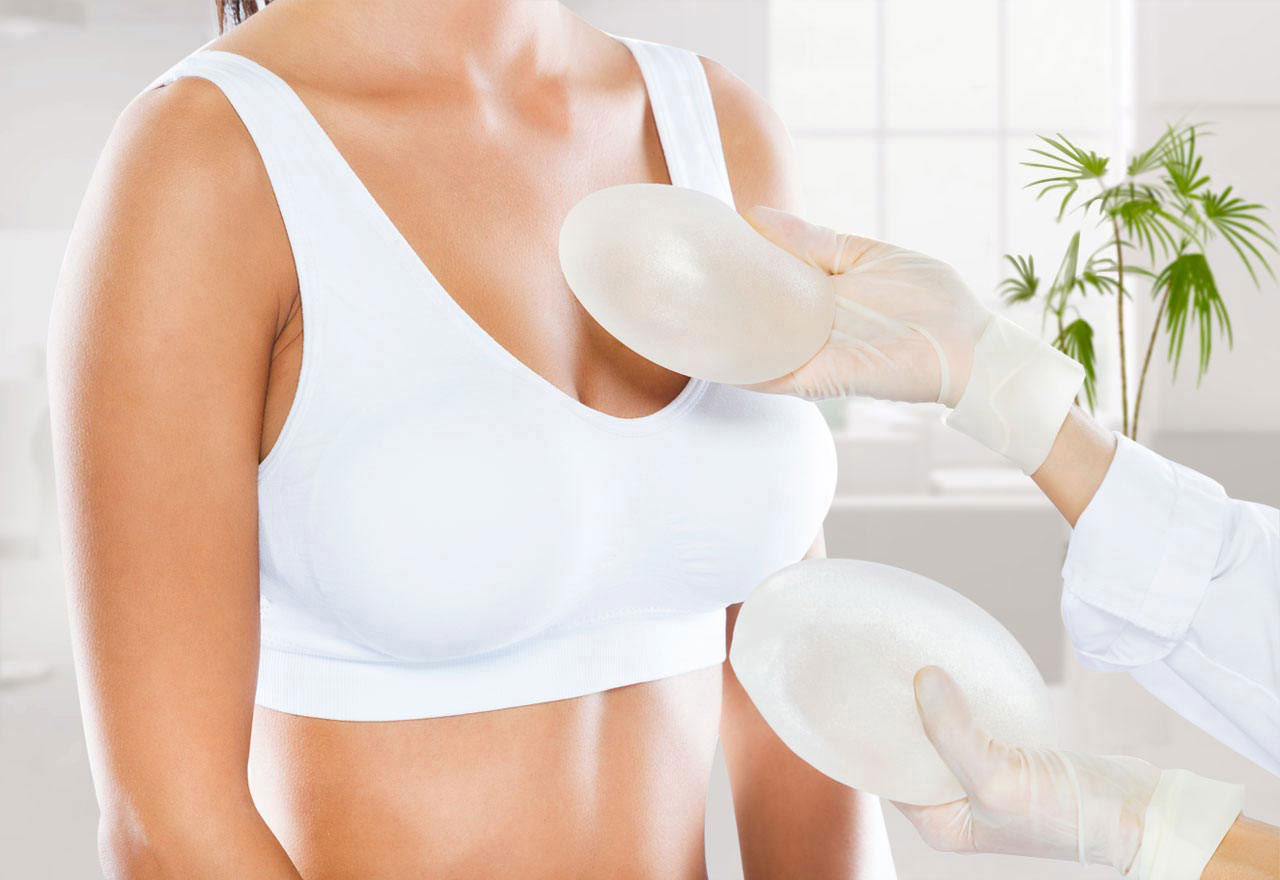
Breast reconstruction surgery aims to reconstruct one or both breasts following a mastectomy (wide local excision or lumpectomy), either immediately following your mastectomy or later. There are two primary forms of reconstruction procedures – implant-based reconstruction and flap reconstruction.
Implant-based reconstruction uses silicone gel or salt water-filled implants to form your new breast mound, making this type of reconstruction quicker and simpler than other techniques, since no tissue needs to be harvested from other parts of your body. However, implant reconstruction doesn’t feel quite as natural and may be less forgiving if weight fluctuations arise in between surgeries.
Flap reconstruction uses skin, fat, and muscle from another area of your body to form new breasts. It often produces natural-feeling firmer breasts than implant reconstruction and may be more suitable for women who do not qualify for implants. If necessary, surgery can also be used to take muscle from either your back (latissimus dorsi muscle reconstruction) or abdomen (DIEP flap reconstruction).
Reconstructive microsurgery techniques involve moving only skin and fat, maintaining blood supply to the original location. Although this procedure could potentially cause arm weakness, this complication is unlikely.
Removal of a Tissue Sample
If you have a lump, your healthcare provider may need to remove a small piece of the tissue to test for cancer. This procedure is known as a biopsy and typically performed under local anaesthesia; occasionally general anaesthesia may be necessary. You will lie on a padded table under an imaging machine known as an MRI (magnetic resonance imaging).
Your healthcare provider will administer local anaesthetic to numb the area around your breast or lump, before performing either a core needle biopsy or vacuum-assisted core biopsy. With either method, they’ll insert a hollow needle into the lump to extract cells for testing; then using special markers they mark where they extracted samples. In both instances a vacuum-assisted core biopsy uses a slightly larger needle attached to an attachment device in order to suction multiple samples at one spot using suction technology – either ultrasound or mammography can assist with guidance; while an ultrasonography-guided core biopsy requires additional guidance based on imaging; both can also use imaging technologies for guidance when performing these types of exams.
If your healthcare provider can’t locate or feel a lump during their examination of you or if its size appears different than expected, they might perform a wire localization procedure in advance of biopsies to locate it using thin metal wire. If this proves inconclusive, surgery might be required to further remove suspicious tissue as well as surrounding tissues that might need removing; then stitches or staples might need to be added at closure – these will typically dissolve with time but some may need to be removed later on.
Drainage Tubes
Surgical drains help drain excess fluid that accumulates after breast tissue has been extracted, potentially forming seromas (see the FAQ page) which cause swelling, discomfort and bruising. Drains prevent this build-up of fluid to speed recovery post surgery.
Drains are pencil-thin flexible tubes that extend into the surgical site with soft plastic bulb at their end to collect excess fluids. Drains can be easily emptied, measured and reset as required; depending on the nature of an operation a person may require one or multiple drains.
Your nurse will brief you during surgery on how to care for and use drains properly, including how to drain tubes properly and log how much drainage there is. After returning home, this practice can continue on its own.
Milking the tube (see FAQ page) can also help clear out your drain. To do this, grasp your tubing close to you body and firmly squeeze while pulling downward towards the drain bulb – this process should be repeated until any clots clogging the tubing have been eliminated; similar to when extracting splinters or foreign objects from skin surfaces.
Pain
pain after breast surgery can be acute and should gradually subside after six to eight weeks. After the operation, you’ll often wear a bandage or dressing over your wound to protect from infection and promote healing. Furthermore, drains – small tubes designed to collect blood or fluid in bags – may remain until healing starts to take place; typically their drainage will decrease within two to five days.
Swelling after surgery is common and can affect your breast, chest wall, shoulder and arm. This condition, called oedema, usually improves with time; it may feel heavy but wearing a supportive bra may help alleviate some discomfort and ease any excess. Always check in with your treatment team if your swelling seems extreme.
Some patients experience pins and needles, itching, or throbbing sensations in their breast and nipple area after having undergone breast reconstruction surgery. This type of pain known as neuropathic discomfort is common and typically lasts several months before subsiding.
Pain levels following breast surgery will depend on both its type and extent; those undergoing an invasive mastectomy (with lymph node removal) are likely to experience longer term discomfort, while reconstruction often increases pain levels further. It’s essential that good pain management strategies be in place even if reconstruction is not in your plans; taking regular doses of ibuprofen can help decrease levels of discomfort while using pain pumps can provide even further pain relief as they “drip” slowly into breast areas when necessary over 2-3 days.
Disclaimer: The content on this blog is intended for general informational purposes only. It is not a substitute for professional medical advice, diagnosis, or treatment. Always consult qualified healthcare providers for personalized advice. Information regarding plastic surgery, dental treatment, hair transplant, and other medical procedures is educational and not a guarantee of results. We do not assume liability for actions taken based on blog content. Medical knowledge evolves; verify information and consult professionals. External links do not imply endorsement. By using this blog, you agree to these terms.


