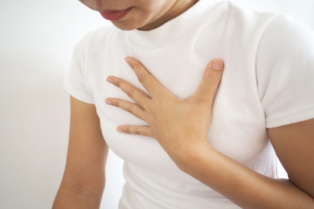
Pectoral muscles are an indispensable muscle group for every bodybuilder. Strengthening them will allow you to press more weight, making both arms stronger in turn.
Most people train their chest using basic exercises such as push ups and chest presses; however, it is also beneficial to incorporate various kinds of chest exercises into their workout regimen so as to ensure even growth of their muscles.
The Ribs
Ribs are long, curved bones that serve to protect vital organs like the heart and lungs from injury while creating space for breathing. Part of your axial skeleton, they connect to sternums and spinal vertebrae in your back for attachment to form the bony thoracic cage; and are divided into three groups – true, false and floating.
True ribs are arranged in pairs and each pair articulates with a specific thoracic vertebrae. Rib 1 connects to T1 vertebrae while subsequent ones correspond with higher vertebrae (for instance rib 2 articulates with T2). Their bodies are thin and flat; their superior surface features blood vessel grooves while their posterior surfaces feature necks with tubercles attached by costotransverse ligaments; these tubercles contain small facet for attachment to transverse processes of their respective vertebrae; tubercles on their posterior surfaces bear small facets for jointing transverse processes of their respective thoracic vertebrae that allows them to articulate with one another thoracic vertebrae pair.
Each rib has an anterior surface that curves inward toward the diaphragm and ends in a cartilage made of hyaline cartilage that may extend several inches from its edge. Most ribs feature attachment sites for deep back muscles; two costal grooves provide space for blood vessels.
The ribs provide space for the lungs to expand during inspiration, helping push air into the chest cavity. Led by external intercostal muscles and diaphragm movement, they are levers used to drive this expansion process. Everturing outward through what’s known as bucket handle motion enables them to increase anteroposterior diameter even further; upper ribs push against the sternum while lower ones evert using costovertebral joints gliding action allows even further anteroposterior diameter increases anteroposterior diameter further by evertracking outward through anteroposterior dilation of thoracic cavity increases anteroposterior diameter further than before enabling greater anteroposterior diameter than ever before!
The Chest Wall
The chest wall serves as a protective cage around the heart, major blood vessels, lungs and liver. Composed of ribs, spine and sternum with muscles and blood vessels enclosing it for support as well as flexibility to allow movement of shoulders and arms, it supports breathing but provides for flexible framework that enables shoulder-arm movement.
The ribs articulate with each other and the sternum through costal cartilages. There are 12 pairs of ribs which connect directly with one another or through costal cartilages to form three groups: (1) Apical Ribs are those which articulate directly with vertebrae; (2) Intermediate Ribs connect directly between themselves and the sternum; and (3) Lateral Ribs do not articulate with either of these entities – all attached directly to spine through costal cartilages with innervated intercostal nerves innervating each rib
There are two openings in the chest cavity: the superior thoracic aperture in front, and inferior thoracic aperture at back, each located along their respective lines of division: superior at front; inferior behind back (bounded by manubrium of sternum, first pair of ribs and body of vertebra T1). Both apertures are covered by diaphragm which serves to divide them both from each other.
The chest (thoracic) wall consists of skin, fat and muscles which form an intricate web of connective tissues to protect vital organs in the chest and upper abdomen. This skeletal framework protects organs such as the heart, aorta and lungs while remaining flexible enough for breathing to take place effectively.
Chest wall disorders may arise for various reasons, including cancerous and noncancerous tumors, infections and trauma. Furthermore, conditions like rheumatoid arthritis of the sternoclavicular joint or systemic lupus erythematosus (SLE) can cause discomfort through inflammation of rib muscles, cartilage or ligaments.
The thoracic skeleton is supported by pectoralis major, latissimus dorsi, serratus anterior and other muscles that form an active support structure for breathing and movement in shoulders and arms. Based on their functional properties these muscles can be divided into inspiratory and expiratory groups depending on when their function takes effect: inspiratory groups expand chest volume during inspiration while expiratory muscles constrict rib cage during exhalation.
The Heart
Your heart is an organ roughly the size of your closed fist that resides at the left side of your chest, pumping blood around to deliver oxygen and nutrients to all the tissues and cells as well as waste products like carbon dioxide from your system.
The heart is a pump consisting of four chambers called atria and ventricles connected by one-way valves; its right and left sides are divided by a wall known as the septum, while each atria connects directly with its respective ventricle via one-way valves. These four heart valves open and close to control blood flow into and out of the heart with each beat, known as the tricuspid valve, mitral valve, aortic valve, and pulmonary valve. These valves function similarly to one-way valves in plumbing systems, preventing blood from flowing back in an unintended direction. As soon as an atria fills with blood, it pushes it out through the tricuspid and mitral valves into both right and left ventricles for circulation. Systole is the initial phase of heartbeats. When the ventricles contract and pump oxygen-rich blood through to the aorta and pulmonary artery, valves open to allow this flow, creating the second sound (dub) associated with heartbeats.
Lungs provide our bodies with oxygen while at the same time producing carbon dioxide, with venous blood returning through large veins called the pulmonary artery and veins to the left atrium for processing before being sent back out through our left ventricle and distributed throughout our bodies.
Your heart muscles must contract at exactly the right time and force, coordinated by electrical impulses from your brain. If it cannot pump effectively, fluid may build up in other parts of the body – particularly lungs, liver and gastrointestinal tract as well as arms and legs; this condition is called congestive heart failure and its cause is usually coronary artery disease where small blood vessels that supply your heart become narrow or blocked, decreasing its ability to pump effectively.
The Lungs
Humans, mammals (and snails) alike all possess two lungs that reside either side of the heart. These organs serve as the heartbeat for your respiratory system by extracting oxygen from air and transporting it throughout your body while exhalation releases carbon dioxide back into the atmosphere.
Every lung is protected by a thin membrane known as the pleura, with capillaries filled with serous pleural fluid providing lubrication to allow smooth breathing and lung expansion/contraction during breathing and change in shape of shape over time. This layer also prevents adhering between lung segments or sticking them against each other or against their respective ribcages, helping the lung expand smoothly as you breathe in or out.
Air enters the lungs via their windpipe, the trachea, which resembles an upside-down “Y.” Once inside, air is warmed, humidified, and cleansed by conducting zones in the lungs – tubes that look like giant sponges which branch off into smaller tubular structures called bronchi. From there they branch off further to form even smaller tubes called bronchioles that branch further still to form small air sacs known as alveoli, each containing fine mesh blood vessels that deliver oxygen while carrying away carbon dioxide.
Any dust that enters these alveoli is attacked by special cells called cilia that cover the surface of lungs’ passageways and brush away any particles that reach them. Cilia also trigger a reflex called Hering-Breuer that protects against overinflation of lungs, and are known for protecting them.
The lungs are roughly cone-shaped organs with an apex, base and three surfaces facing towards the ribs. Since one lung shares space with the heart, its left lung features an indentation called a cardiac notch to accommodate this fact.
The lungs are divided into lobes – each lung typically contains two or three, further subdivided by septa. Every lung features surfaces facing towards its respective ribcage, diaphragm and surface between that is known as an oblique fissure – with its apex pointing upward towards ceiling while its base lies flat against ribs – separated from pulmonary hilum by this fissure.
Disclaimer: The content on this blog is intended for general informational purposes only. It is not a substitute for professional medical advice, diagnosis, or treatment. Always consult qualified healthcare providers for personalized advice. Information regarding plastic surgery, dental treatment, hair transplant, and other medical procedures is educational and not a guarantee of results. We do not assume liability for actions taken based on blog content. Medical knowledge evolves; verify information and consult professionals. External links do not imply endorsement. By using this blog, you agree to these terms.


